Pneumonia Detection from Chest X-Rays
The Unseen Crisis in Pneumonia Diagnosis
Pneumonia remains a significant health challenge, particularly in developing and underdeveloped nations. The disease, caused by bacterial or viral infections, leads to pleural effusion, where fluids fill the lungs, causing severe respiratory difficulties. Early diagnosis is crucial for effective treatment and improving survival rates. However, the examination of chest X-rays, the most common diagnostic method, is fraught with challenges and subjective variability.

The Limitations of Traditional Diagnostic Methods
Traditional machine learning algorithms have been employed for pneumonia detection, but they require extensive domain expertise for feature extraction. This process is not only time-consuming but also prone to human error. Moreover, the lack of publicly available data has been a significant limitation, hindering the development of robust diagnostic models.
The Role of Deep Learning in Pneumonia Detection
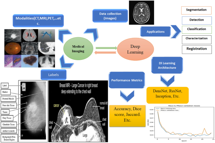
Deep learning models have revolutionized the field of medical imaging, offering a variety of architectures such as VGGNet, ResNet, and Inception ResNet. These models, when combined with transfer learning techniques, have shown promise in improving diagnostic accuracy. However, the challenge remains in effectively extracting and utilizing the region of interest in chest X-rays to identify pneumonia.

Real Case Studies: Successes and Shortcomings
Several studies have attempted to address these challenges with varying degrees of success. For instance, a study using ResNet18 on a small dataset of 349 chest X-ray images achieved an impressive accuracy of 99.4%. However, the limited dataset size raises concerns about the model’s generalizability. Another study employed an ensemble of deep learning models, including GoogLeNet, ResNet-18, and DenseNet-121, achieving high accuracy rates on two publicly available datasets. Despite these successes, the reliance on experimental methods for weight assignment in ensemble models introduces potential errors.
Our Solution: Integrating Computer Vision + GPT-4o
In our AI in Medical course, we teach a comprehensive solution for pneumonia detection in chest X-rays. Our approach involves training a ResNet50 classifier integrated with an LLM-based assistant to provide auxiliary disease information. This integration enhances the model’s diagnostic capabilities by leveraging additional contextual data. The user-friendly Gradio-chat interface allows for seamless X-ray uploads, making the tool accessible to healthcare professionals. We provide full code access, a detailed tutorial, and an explanatory presentation to ensure a thorough understanding of the solution.
Learn to Code This Inside Out
You can master this advanced AI solution in our AI in Medical Course. For just $59, you will gain access to all the resources needed to implement this cutting-edge technology. This offer ends tonight, so don’t miss out on the opportunity to enhance your skills and contribute to the future of medical diagnostics.
If you found this article interesting and want to be on the cutting edge of AI, then ready yourself to level up your AI and Computer Vision skills with practical, industry-relevant knowledge. Join us at Augmented AI University and bridge the gap between academic learning and the skills you need in the workplace.
Don’t miss out on the opportunity to enhance your career with cutting-edge AI education. Enroll now and start building a foundation of practical AI skills to tackle tomorrow’s technological challenges!
Enroll in Augmented AI University Today!

